|
Spicules of the coccyx - 3 / Epines du coccyx - 3
Jean-Yves Maigne, MD
|
| |
|
Spicules;
cases #15 to 20
|
| |
 When the tip
of the coccyx is hardly visible on the radiographs, a CT or an MRI scan are two
excellent options. See below. When the tip
of the coccyx is hardly visible on the radiographs, a CT or an MRI scan are two
excellent options. See below.
 Si la
pointe du coccyx est mal visible sur les radios, le scanner ou l'IRM sont deux
examens interressants. Voir ci-dessous. Si la
pointe du coccyx est mal visible sur les radios, le scanner ou l'IRM sont deux
examens interressants. Voir ci-dessous.
|
|
 Case #15, 16:
Two different cases (lateral and posterior views). The spicule is very
different in nature from an osteophyte, which is in fact a bony ring
(i.e. circular). Case #15, 16:
Two different cases (lateral and posterior views). The spicule is very
different in nature from an osteophyte, which is in fact a bony ring
(i.e. circular).
 Cas 15 et
16. Deux cas différents (vues de profil et de face). Le spicule
est de nature diférente d'un ostéophyte, qui n'est qu'une collerette
osseuse. Cas 15 et
16. Deux cas différents (vues de profil et de face). Le spicule
est de nature diférente d'un ostéophyte, qui n'est qu'une collerette
osseuse. |
|
|
| |
|
|
 Case #20:
This lady felt like she was sitting on a "bunch of pebbles". The X-rays
were quite normal, but MRI demonstrated an hypertrophic distal piece of
bone turned backward and downward, i.e. to the skin (look at the picture
on the right). An anesthetic block test has been positive (she didn't
want any cortisone injection), and she was operated on, with a good
result. Case #20:
This lady felt like she was sitting on a "bunch of pebbles". The X-rays
were quite normal, but MRI demonstrated an hypertrophic distal piece of
bone turned backward and downward, i.e. to the skin (look at the picture
on the right). An anesthetic block test has been positive (she didn't
want any cortisone injection), and she was operated on, with a good
result.
 Cas 20.
Cette femme avait l'impression d'être assise sur un sac de
cailloux. Les radios étaient quasiment normales, mais l'IRM mit en
évidence une pièce osseuse distale hypertrophiée orientée en bas et en
arrière, vers la peau. Le bloc anesthésique ayant été positif, elle fut
opérée avec un bon résultat. Cas 20.
Cette femme avait l'impression d'être assise sur un sac de
cailloux. Les radios étaient quasiment normales, mais l'IRM mit en
évidence une pièce osseuse distale hypertrophiée orientée en bas et en
arrière, vers la peau. Le bloc anesthésique ayant été positif, elle fut
opérée avec un bon résultat. |
 |
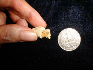
 She
cleaned up her coccyx and took this picture. The spicule is pointing
downward. The surgeon performed a total coccygectomy, but in my opinion,
a partial one would have been successful as well. She
cleaned up her coccyx and took this picture. The spicule is pointing
downward. The surgeon performed a total coccygectomy, but in my opinion,
a partial one would have been successful as well.
 Le
coccyx a été nettoyé et pris en photo. Le spicule est visible, pointant
vers le bas. Ici, une coccygectomie partielle aurait sufit, à mon avis. Le
coccyx a été nettoyé et pris en photo. Le spicule est visible, pointant
vers le bas. Ici, une coccygectomie partielle aurait sufit, à mon avis. |
| |
|
Comment réaliser et lire les radiographies dynamiques
1 |
|
Luxations
postérieures 1 -
2 |
|
Hypermobilité
1 -
2 |
| Epines
1 -
2 -
3 |
|
Luxations antérieures 1 |
|
Radiographies "normales" |
| Lésions
complexes 1 |
|
Fractures
1 |
|
Calcifications
1 -
2 |
|
Déformations
1 |
|
Anatomie du coccyx |
|
