|
Hypermobility
of the coccyx -2
Jean-Yves Maigne, MD
|
Here are seven
other cases of hyper mobility. You will learn about another radiologic sign: the
misalignment of the coccygeal vertebrae (also called "bones") in the sitting
position, which is an accompanying sign of hyper mobility in flexion, and you
will see two examples of a frequent difficulty in reading the radiographs, when
the distal part of the coccyx is involved.
| |
|
Seven other cases of hypermobility
|
| |
|
|
|
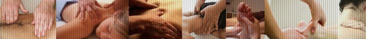
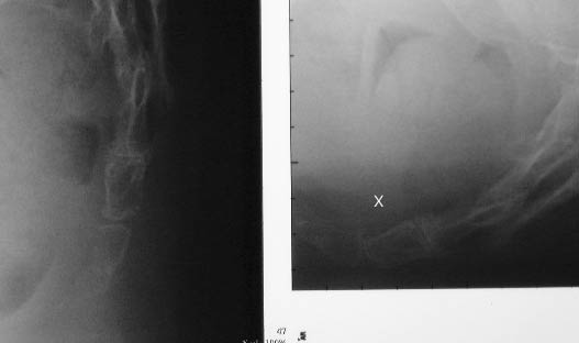
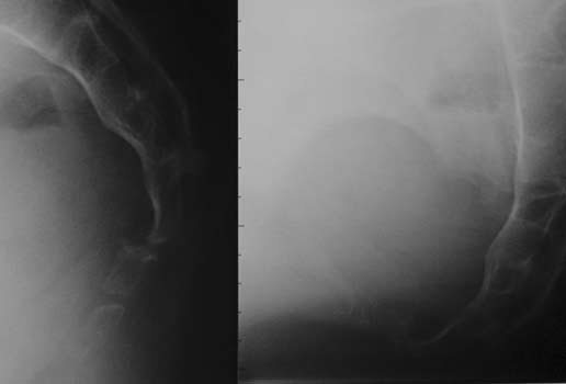
Case #7. Hyper mobility can also be accompanied by another
interesting sign: a misalignment of the two involved bones (a step in
their anterior aspect) which is obvious in this case, where the two
other radiologic signs are present : hyper mobility (40į) and bony
contact with friction. |
|
|
|
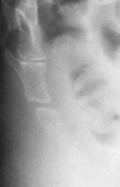
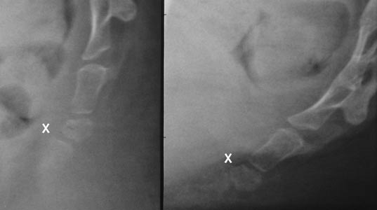
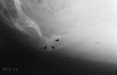
Case #8. The mobility is between 25į and 30į, but there is a
friction of the bones and a misalignment in flexion, which I consider
abnormal (despite very slight here). Naturally, the interspace was
tender at palpation. |
|
|
|
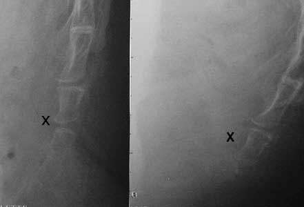
Case #9. Another example of misalignment in flexion (X). |
|
|
|
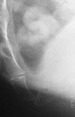
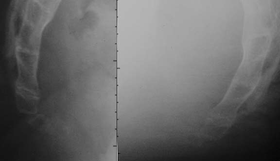
Case #10. A misleading aspect. The distal bone, not clearly
visible on the sitting film here, has pivoted of 50į in flexion. The
patient felt her pain at the tip of the coccyx, a vey good clinical
indicator. |
|
|
|
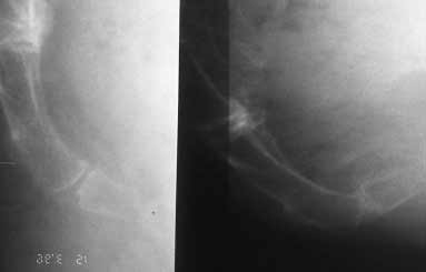
Case #11. Another case with hyper mobility and friction (note
on the sitting film how the opposite surfaces are congruent) but without
misalignment. |
|
|
|
Case #12. this case is similar to case #9, the distal part of
the coccyx being difficult to evidence on the film. |
|
|
|
Case #13. A strange aspect. There is clearly an hyper mobility,
but the coccyx seems to be in a permanent state of anterior luxation.
|
|
|
|
Case #14. This is a "sitting film" with a marked misalignment
and an irregularity of the endplate, with a step, maybe corresponding to
the sacral apex. |
|
Comment rťaliser et lire les radiographies dynamiques
1 |
|
Luxations
postťrieures 1 -
2 |
|
Hypermobilitť
1 -
2 |
| Epines
1 -
2 -
3 |
|
Luxations antťrieures 1 |
|
Radiographies "normales" |
| Lťsions
complexes 1 |
|
Fractures
1 |
|
Calcifications
1 -
2 |
|
Dťformations
1 |
|
Anatomie du coccyx |
|