|
Deformities of the coccyx / Dťformations du coccyx
Jean-Yves Maigne, MD
|
 Deformities of the
coccyx are rare. They may affect its axis in the frontal plane (lateral
deviation), its shape or its lenght. Deformities of the
coccyx are rare. They may affect its axis in the frontal plane (lateral
deviation), its shape or its lenght.
 Les dťformations du
coccyx sont rares. Elles concernent son axe dans le plan frontal (dťviation
latťrale), sa forme ou sa longueur.
Les dťformations du
coccyx sont rares. Elles concernent son axe dans le plan frontal (dťviation
latťrale), sa forme ou sa longueur.
| |
|
Deviation in the frontal
plane / Dťviations frontales
|
| |
|
|
|
Case
#1: A slight frontal deviation is often the continuation of a
lumbar scoliosis. See also fig 4 of the paper by Kim and Suk at
http://www.coccyx.org/medabs/kimsuk.htm
Cas
1 : Une dťviation frontale est souvent la continuation d'une
scoliose lombaire. Voir aussi la fig 4 de l'article de Kim & Suk ŗ
http://www.coccyx.org/medabs/kimsuk.htm |
|
|
|
Case
#2: The deviation may also be isolated without lumbar
scoliosis. Relationship with pain is unclear
Cas
1 : la dťviation peut aussi Ítre isolťe sans scoliose lombaire.
Les rapports avec la douleurs sont peu clairs |
|
|
|
Case
#3: The deviation is likely to be post traumatic in this
particular case
Cas
1 : Ici, la dťviation est probablement post-traumatique |
| |
|
Abnormal shape (in
the sagittal plane) / Forme anormale de profil
|
| |
|
|
|
|
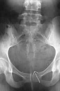
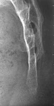
Case
#1, 2: The thickening of the distal part of the coccyx is in
fact a rare variety of "giant" spicule pressing or crushing the
subcutaneous tissues in the sitting position. The coccyx is "club
shaped". The joints are fused and the coccyx is deprived of any
mobility. All the cases I have observed were accompanied by a cutaneous
pit or a pilonidal sinus. Case #1, on the left, is a very moderate case.
Case #2 is self speaking.
Cas
1 : L'ťpaississement de la partie distale du coccyx est en fait
une variťtť rare d'ťpine gťante ťcrasant les tissus sous cutanťs en
station assise. Le coccyx est en forme de massue, les articulations sont
fusionnťes, supprimant toute mobilitť. Tous les cas que j'ai observťs
ťtaient accompagnťs d'une fossette cutanťe ou d'un sinus pilonidal. A
gauche, un cas modťrť. A droite, sans commentaire. |
|
|
|
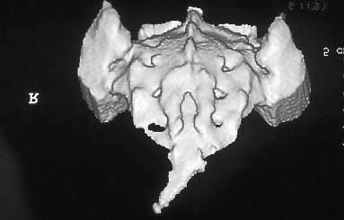
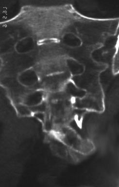
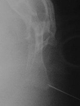
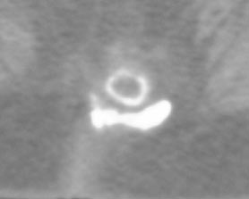
Case #3:
This is a
very abnormal giant spicule, which co-existed with a pilonidal sinus.
The CT shows the curious shape of this excrescence, dorsal and lateral.
Surgery has been offered to the patient, with a good result.
Cas
1 : un spicule gťant trŤs atypique, associť ŗ un sinus
pilonidal. Le scanner montre la curieuse forme de l'excroissance,
dorsale et latťrale. La patiente a ťtť opťrťe avec un bon rťsultat. |
| |
|
Abnormal coccygeal
lenght / Anomalies de longueur
|
| |
|
|
|
|
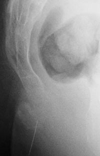
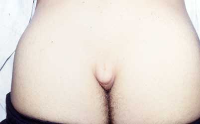
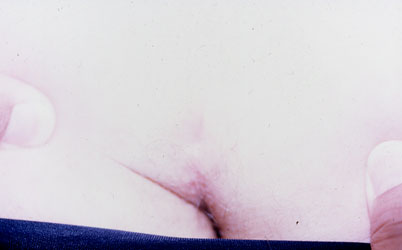
Case
#1, 2: Two cases where the coccyx was too long, like a human
tail. The handicap is not only cosmetic, but functional, making
prolonged sitting positions very uncomfortable. See also a very similar
case at
http://www.coccyx.org/personal/2003/anon0317.htm
Cas
1 et 2: deux cas de coccyx trop long, comparable ŗ une queue
humaine. Le handicap n'est pas seulement esthťtique mais fonctionnel,
rendant les stations assises prolongťes trŤs inconfortables. Voyez un
cas similaire. |
|
Comment rťaliser et lire les radiographies dynamiques
1 |
|
Luxations
postťrieures 1 -
2 |
|
Hypermobilitť
1 -
2 |
| Epines
1 -
2 -
3 |
|
Luxations antťrieures 1 |
|
Radiographies "normales" |
| Lťsions
complexes 1 |
|
Fractures
1 |
|
Calcifications
1 -
2 |
|
Dťformations
1 |
|
Anatomie du coccyx |
|