image
image
image
image


Calcifications of the coccyx - 2

Jean-Yves Maigne, MD


Below are 
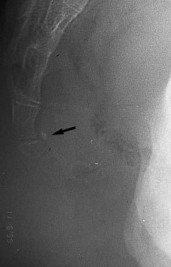
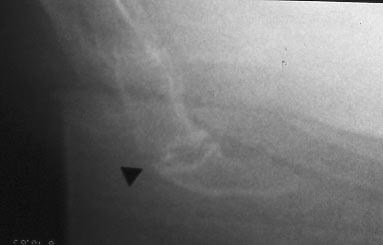
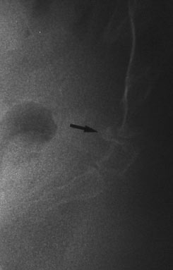
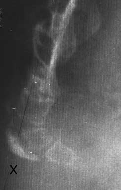
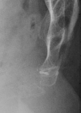
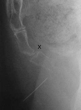
some more common cases of calcifications of the coccygeal discs.

Voici quelques cas plus habituels de calcifications coccygiennes.

 
cases #6 to 11
 
Case #6: permanent calcification of the intercoccygeal disc
Cas 6 : calcification permanente du disque intercoccygien
Case #7: intradiscal calcification
Cas 7 : calcification intra-discale
 
 
Case #8: anterior calcification
Cas 8 : Calcification antérieure
Case #9: A strange image. I presume it is a calcified hematoma, but I am not sure
Cas 9 : une curieuse image. Je pense qu'il s'agit d'un hématome calcifié, mais sans certitude
Case #10 and 11 : Intradiscal calcification. In both cases, the lower disc was the source of pain, as the success of an injection can testify
Cas 10 et 11: calcifications intra-discales. Dans les deux cas, c'est le disque sous-jacent qui était la cause de la douleur, come en témoigne le succès de l'injection intra-discale
 
Content
 
Comment réaliser et lire les radiographies dynamiques 1
Luxations postérieures 1 - 2
Hypermobilité 1 - 2
Epines 1 - 2 - 3
Luxations antérieures 1
Radiographies "normales"
Lésions complexes 1
Fractures 1
Calcifications 1 - 2
Déformations 1
Anatomie du coccyx


image
 
image
image
image