|
Rameaux dorsaux
thoraco-lombaires
Thoracolumbar dorsal rami
Trajet profond
/ Deep course (1)
|

 La racine spinale
thoracique basse et ses branches de division : le rameau ventral en
avant (nerf intercostal) et le rameau dorsal en arrière, très court et se
divisant immédiatement en une branche latérale (musculaire et cutanée, donc
épaisse) et une branche médiale (innervant l'arc postérieur et le multifidus),
plus mince. La racine spinale
thoracique basse et ses branches de division : le rameau ventral en
avant (nerf intercostal) et le rameau dorsal en arrière, très court et se
divisant immédiatement en une branche latérale (musculaire et cutanée, donc
épaisse) et une branche médiale (innervant l'arc postérieur et le multifidus),
plus mince.
 The lower thoracic
spinal root and its branches: the ventral ramus (intercostal nerve) and
the dorsal ramus, very short and immediately dividing into a lateral branch
(muscular and cutaneous, thus thick) and a tiny medial branch, which supplies
the dorsal arch and the multifidus. The lower thoracic
spinal root and its branches: the ventral ramus (intercostal nerve) and
the dorsal ramus, very short and immediately dividing into a lateral branch
(muscular and cutaneous, thus thick) and a tiny medial branch, which supplies
the dorsal arch and the multifidus.
 |
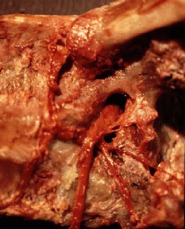
 Vue latérale du
foramen T11-12 gauche. En haut, la 11ème côte, en avant, les
deux corps vertébraux. La racine sort du foramen intervertébral pour
former le nerf intercostal. Le rameau dorsal et ses deux branches sont
bien visibles, le rameau médial passant au pied de l'articulation
zygapophysaire T11-12, dont la capsule a été incisée. Vers l'avant part
le rameau communiquant destiné à la chaine sympathique. Vue latérale du
foramen T11-12 gauche. En haut, la 11ème côte, en avant, les
deux corps vertébraux. La racine sort du foramen intervertébral pour
former le nerf intercostal. Le rameau dorsal et ses deux branches sont
bien visibles, le rameau médial passant au pied de l'articulation
zygapophysaire T11-12, dont la capsule a été incisée. Vers l'avant part
le rameau communiquant destiné à la chaine sympathique.
 Lateral
view of the left T11-12 foramen. At the top, the 11th rib and
on the left of the photo, the two vertebral bodies. The root exits the
intervertebral foramen and becomes the intercostal nerve. The dorsal
ramus and its two branches are well visible, the medial branch running
at the foot of theT11-12 zygapophyseal joint, the capsule of which
having been severed. The ramus comminicansruns ventrally to the
sympathetic trunk. Lateral
view of the left T11-12 foramen. At the top, the 11th rib and
on the left of the photo, the two vertebral bodies. The root exits the
intervertebral foramen and becomes the intercostal nerve. The dorsal
ramus and its two branches are well visible, the medial branch running
at the foot of theT11-12 zygapophyseal joint, the capsule of which
having been severed. The ramus comminicansruns ventrally to the
sympathetic trunk. |
 |
 Une vue
similaire d'une autre dissection. La flèche montre la branche
postérieure avant sa division. T11 et T12 marquent le corps vertébral.
Le disque T12-L1 (fibres blanches verticales) apparait très épais. Ceci
est dû au fait que les fibres annulaires les plus latérales ne
s'attachent pas sur les plateaux vertébraux, mais s'insèrent sur la
partie latérale des corps vertébraux. Une vue
similaire d'une autre dissection. La flèche montre la branche
postérieure avant sa division. T11 et T12 marquent le corps vertébral.
Le disque T12-L1 (fibres blanches verticales) apparait très épais. Ceci
est dû au fait que les fibres annulaires les plus latérales ne
s'attachent pas sur les plateaux vertébraux, mais s'insèrent sur la
partie latérale des corps vertébraux.
 A similar
view of another dissection. The arrow shows the dorsal ramus
before its division. T11 and T12 indicate the vertebral body. The T12-L1
disc (white vertical fibers) appears very thick. This is due to the fact
that the most lateral annular fibers insert not on the endplates, but on
the lateral aspect of the vertebral bodies. A similar
view of another dissection. The arrow shows the dorsal ramus
before its division. T11 and T12 indicate the vertebral body. The T12-L1
disc (white vertical fibers) appears very thick. This is due to the fact
that the most lateral annular fibers insert not on the endplates, but on
the lateral aspect of the vertebral bodies. |
Contenu / Content
| ©2003 Jean-Yves Maigne,
MD
Accueil Anatomie • Home Page Anatomy |
Trajet profond / Deep course |
1 • 2 |
| Trajet
superficiel / Superficial course |
1 •
2 • 3 |
| Croisement de
la crête iliaque / Crossing the iliac crest |
1 •
2 |
|
Territoire d'innervation / Territory of distribution |
1 •
2 |
|
Anastomoses / Anastomosis |
1 |
|
