
|
|
|
|
|
Rameaux dorsaux thoraco-lombaires
Thoracolumbar dorsal rami
Introduction |
Contenu / Content
| ©2003 Jean-Yves Maigne, MD
Accueil Anatomie • Home Page Anatomy |
Trajet profond / Deep course |
1 •
2 |
| Trajet superficiel
/ Superficial course |
1 •
2 • 3 |
| Croisement de la
cręte iliaque / Crossing the iliac crest |
1 •
2 |
|
Territoire d'innervation / Territory of distribution |
1 •
2 |
|
Anastomoses / Anastomosis |
1 |
|
Introduction aux dissections des rameaux dorsaux
Nous nous sommes intéressés aux branches postérieures issues de la
charničre thoraco-lombaire suite aux études de Robert
Maigne sur les lombalgies provenant d'un dysfonctionnement de cette
région. Cet auteur avait remarqué que certaines douleurs lombaires
basses étaient en fait des douleurs projetées ŕ partir de la charničre
thoraco-lombaire et il avait pratiqué quelques dissections de ces
rameaux dorsaux. Il en avait conclu que la carte proposée par Keegan et
Garett (voir ci-dessous) ne correspondait pas ŕ la réalité. La région
fessičre était bien innervée par des rameaux issus des racines T11, T12
et L1 et non par ceux provenant de L4, L5 ou S1. En 1990, nous avons
entrepris une étude sur 37 dissections, ce qui nous a permis de
confirmer cette notion, et de mettre en évidence la possibilité d'un
syndrome canalaire lorsque le rameau nerveux le plus interne croisait la
cręte iliaque. Ce sont ces dissections que nous présentons ici.
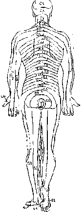
 La célčbre
carte des dermatomes de Keegan et Garett. Nos résultats ne
confirment pas cette carte en ce qui concerne la région lombaire
basse. Ils sont résumés sur les trois schémas ci-dessous (travail
sur 37 dissections). La célčbre
carte des dermatomes de Keegan et Garett. Nos résultats ne
confirment pas cette carte en ce qui concerne la région lombaire
basse. Ils sont résumés sur les trois schémas ci-dessous (travail
sur 37 dissections).
 The
famous dermatome chart by Keegan and Garett. Our results
are not in accordance with this chart, as far as the lower back is
concerned. They are summarised on the three sketches below (study on
37 dissections). The
famous dermatome chart by Keegan and Garett. Our results
are not in accordance with this chart, as far as the lower back is
concerned. They are summarised on the three sketches below (study on
37 dissections).
|

Introduction to
the
dissections of the dorsal rami of the thoracolumbar junction
We took an interest
in the dorsal rami from the thoracolumbar junction
after the studies by Robert Maigne on low back pain related to a
dysfunction of that junction. This author had noticed that some low
back pain syndromes were actually referred pain syndromes from the
thoracolumbar junction, and he performed a few dissections of these
rami. His conclusions were that the dermatomal chart by Keegan and
Garett (see below) was not exact. The gluteal area was in fact
supplied by the cutaneous rami arising from the T11, 12 and L1
spinal roots, and not from L4, 5 or S1. In 1990, we undertook an
anatomic study on 37 dissections, which allowed us to confirm this
fact, and to demonstrate the possibility of an entrapment
neuropathy, where the most medial nerve crossed the iliac crest.
These dissections are presented in this section.
|
|
Our results were published
in:
The lateral cutaneous branches of the dorsal rami of the
thoracolumbar junction. An anatomical study on 37 dissections,
Maigne JY, Lazareth JP, Guerin Surville H, Maigne R. Surg Radiol Anat
1989;11:289-93.
Trigger point of the posterior iliac crest: painful iliolumbar
ligament insertion or cutaneous dorsal ramus pain? An anatomic study,
Maigne JY, Maigne R.
Archives of Physical Medicine and Rehabilitation. 1991;72:734-7.
|
 For presentation purpose, the course of the nerves is divided in five
parts, from the deep to the superficial layer. Each layer is comprised
of one to three pages of photos. Each page weights around 200 Ko.
For presentation purpose, the course of the nerves is divided in five
parts, from the deep to the superficial layer. Each layer is comprised
of one to three pages of photos. Each page weights around 200 Ko.
|
 |
|
La
carte des dermatomes telles que nos dissections ont pu l'établir.
The actual dermatome chart of the low back, according to the results of
our dissections. |
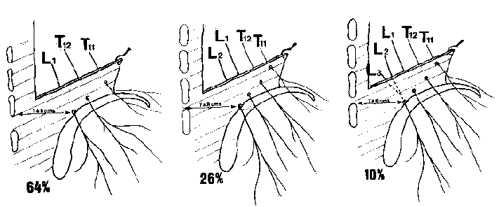
| Disposition la
plus fréquente (64% des cas) : le rameau cutané le plus bas est
L1.
The most frequent pattern (64% of the
cases) is L1 as the lower cutaneous dorsal ramus. |
Dans 26% des cas,
le rameau cutané le plus bas est L2. In
26% of the cases, the lower cutaneous ramus is L2 |
Dans 10% des cas,
le rameau cutané le plus bas est L3, mais nous ne l'avons trouvé qu'en
tant qu'anastomose de L2.
In only 10% of the cases, the lower
ramus is L3, but we only found it as an anastomosis of L2 (and not as an
independant nerve). |
|
Dans
ces trois cas, remarquez que le nerf le plus interne croise la cręte
iliaque ŕ une distance constante qui est d'environ 7 centimčtres de la
ligne médiane. Pour plus de détails, voir les dissections.
Note that in these three cases, the most medial nerve crosses the iliac
crest at a distance of about 7 centimetres from the midline. For more
details, see the dissections. |
|
Articles sur ce sujet /
Articles on the same topic
|
| Maigne R. Low back pain
of thoracolumbar origin.
Archives of Physical Medicine and Rehabilitation.
1980;61:389-395.
Texte disponible ici |
| Maigne JY, Doursounian
L. Entrapment neuropathy of the medial superior cluneal nerve. Nineteen
cases surgically treated, with a minimum of 2 years' follow-up. Spine
1997 May 15;22:1156-9.
Cliquer ici pour le résumé |
| Maigne JY, Maigne R.
Trigger point of the posterior iliac crest: painful iliolumbar ligament
insertion or cutaneous dorsal ramus pain? An anatomic study. Arch Phys
Med Rehabil 1991 Sep;72(10):734-7.
Texte disponible ici |
| Maigne JY, Doursounian
L, Maigne R. [Tunnel syndrome of the dorsal ramus of L1 at the iliac
crest. Treatment by neurolysis] Rev Rhum Mal Osteoartic. 1991;58:230-1.
No abstract available. |
| Maigne JY, Lazareth JP,
Guerin Surville H, Maigne R.The lateral cutaneous branches of the dorsal
rami of the thoraco-lumbar junction. An anatomical study on 37
dissections. Surg Radiol Anat 1989;11(4):289-93.
Cliquer ici pour le résumé |
| Maigne JY, Lazareth JP,
Maigne R. [Anatomical study of the cutaneous innervation of the
lumbosacral region. Application to the physiopathology of certain
lumbalgias]. Rev Rhum Mal Osteoartic. 1988;55:107-11. |
|
|
|
|
|
|
|
|
|
|