|
Rameaux
perforants latéraux (T12 et L1)
Lateral perforating rami (T12 and L1)
|
Contenu / Content
| Croisement de
la cręte iliaque / Crossing the iliac crest
|
1 •
2 |
| Distribution / Distribution |
1 |
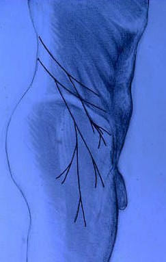
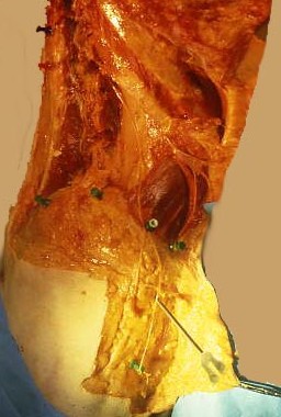
Les rameaux perforants latéraux sont des nerfs
sensitifs issus de la partie latérale des nerfs sous-costal (rameau ventral de
T12) et iliohypogastrique (rameau ventral de L1). Ils croisent la cręte iliaque
latérale et se dirigent vers le grand trochanter. Ils innervent la peau et les
tissus sous-cutanés de la région de la face externe de la hanche. Les traités
classiques les décrivent sous la forme de nerfs courts sans pathologie
spécifique. R. Maigne avait montré que des douleurs provenant de la charničre
thoraco-lombaire pouvaient irradier dans leur territoire d'innervation et
descendre assez bas (jusqu'ŕ mi-cuisse). Notre travail anatomique a confirmé que
ces nerfs pouvaient descendre plus de 10 cm sous le trochanter, et qu'un
syndrome canalaire était possible lorsqu'ils croisaient la cręte iliaque.
 The lateral perforating
rami are both sensory nerves arising from the lateral portion of the subcostal
(ventral ramus of T12) and iliohypogatric (ventral ramus of L1) nerves. They
cross the lateral iliac crest and turn toward the trochanter. They supply the
skin and the subcutaneous tissues of the lateral aspect of the hip area. They
are described in the textbooks as short nerves without any specific pathology.
R. Maigne had shown that pain from the thoracolumbar junction could radiate in
their territory of distribution and extend quite low (to mid-thigh). Our
anatomic study confirmed that these nerves could run up to 10 cm below the
greater trochanter, and that the prerequisites for an entrapment neuropathy were
present when they crossed the iliac crest. The lateral perforating
rami are both sensory nerves arising from the lateral portion of the subcostal
(ventral ramus of T12) and iliohypogatric (ventral ramus of L1) nerves. They
cross the lateral iliac crest and turn toward the trochanter. They supply the
skin and the subcutaneous tissues of the lateral aspect of the hip area. They
are described in the textbooks as short nerves without any specific pathology.
R. Maigne had shown that pain from the thoracolumbar junction could radiate in
their territory of distribution and extend quite low (to mid-thigh). Our
anatomic study confirmed that these nerves could run up to 10 cm below the
greater trochanter, and that the prerequisites for an entrapment neuropathy were
present when they crossed the iliac crest.
Our results were published in:
The lateral cutaneous
branches of the dorsal rami of the thoracolumbar junction. An anatomical study
on 37 dissections,
Maigne JY, Lazareth JP,
Guerin Surville H, Maigne R. Surg Radiol Anat 1989;11:289-93.
|