|

|
|
|
|
Rameaux perforants latéraux : croisement de la cręte iliaque (1)
Lateral perforating rami: crossing the crest (1)
|
Contenu / Content
| Croisement de
la cręte iliaque / Crossing the iliac crest
|
1 • 2 |
| Distribution / Distribution |
1 |
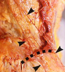 |
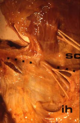
 Vue
postérieure droite des nerfs sous-costal (en haut) et
ilio-hypogastrique (et en bas). Ils abandonnent un rameau perforant ŕ
une distance variable de la cręte iliaque. La séparation est bien
visible et est marquée par les deux flčches noires supérieures. On
notera que le rameau perforant de L1 est plus épais que le nerf
principal (aprčs la séparation), probablement en raison d'un territoire
d'innervation plus étendu. La ligne pointillée marque la cręte iliaque.
Le rameau perforant de L1 la croise ŕ sa partie la plus latérale (ŕ la
verticale du grand trochanter), celui de T12 plus en avant. Vue
postérieure droite des nerfs sous-costal (en haut) et
ilio-hypogastrique (et en bas). Ils abandonnent un rameau perforant ŕ
une distance variable de la cręte iliaque. La séparation est bien
visible et est marquée par les deux flčches noires supérieures. On
notera que le rameau perforant de L1 est plus épais que le nerf
principal (aprčs la séparation), probablement en raison d'un territoire
d'innervation plus étendu. La ligne pointillée marque la cręte iliaque.
Le rameau perforant de L1 la croise ŕ sa partie la plus latérale (ŕ la
verticale du grand trochanter), celui de T12 plus en avant. |
 This is a right posterior view of the the subcostal (above) and
iliohypogastric (below) nerves. They abandon a perforating ramus at a
variable distance from the crest. The separation is clearly visible and
is marked by the upper arrows. Interestingly, the perforating ramus of
L1 is thicker than the primary nerve (considered after the separation),
supposedly because the territory of distribution of the former is
larger. The dotted line shows the iliac crest. The perforating ramus of
L1 crosses it at its most lateral portion, vertically above the
trochanter, the one of T12 more anteriorly.
This is a right posterior view of the the subcostal (above) and
iliohypogastric (below) nerves. They abandon a perforating ramus at a
variable distance from the crest. The separation is clearly visible and
is marked by the upper arrows. Interestingly, the perforating ramus of
L1 is thicker than the primary nerve (considered after the separation),
supposedly because the territory of distribution of the former is
larger. The dotted line shows the iliac crest. The perforating ramus of
L1 crosses it at its most lateral portion, vertically above the
trochanter, the one of T12 more anteriorly. |
|
|
|
|
|
|
|
|
|
|