Written 1997
Thoracolumbar junction
The role of the cutaneous dorsal rami
Jean-Yves Maigne, MD and Robert Maigne, MD, Physical
Medicine, Hotel-Dieu Hospital, 75181 Paris Cedex 04,
France
|
Abstract:
The
thoracolumbar junction (T10-11 to L1-2), and the sensory nerves arising at that
level, may be responsible for pain referred to the lower back. This pain is
distinguished by its topography, in the distribution of the superior cluneal
nerves; by hypersensitivity of the skin and subcutaneous tissues in this territory;
and by a point on the iliac crest, about 7 cm from the midline, pressure on
which produces the pain familiar to the patient. The pain may result from facet
joint dysfunction, rather than from disk disorders. The syndrome is usually
treated with local injections and vertebral manipulation.
The source of low
back pain is usually sought in lesions of the disks or the facet joints at the
L4-L5 and L5-S1 levels. However, in a carefully conducted study, almost 50%
of the patients with low back pain without radiation into the lower limbs had
pain that clearly was neither discogenic nor caused by the facet joints.(19)
Many other structures, such as ligaments, the sacroiliac joints, or trigger
points, have been incriminated. The thoracolumbar junction (TLJ), and the nerves
arising at that level, may be implicated in a major way: The lumbar region receives
its sensory innervation from these nerves; and pain in that region may have
been referred from a higher level in the spine.
|
|
The
thoracolumbar junction is linked
to the lower lumbar region |
| |
The innervation
of the lower lumbar region, below the iliac crest, is provided by sensory nerves
that come from the T11, T12, L1 and L2 roots. At each level, these roots give
rise to a short dorsal ramus, which divides into two branches, a medial one
and a lateral one. These branches course caudal, crossing over the iliac crest
to innervate the skin. They are known as the superior cluneal nerves. This pattern
has been known to anatomists since the end of the 19th century;(3) however,
the concept was superseded by the dermatomal chart drawn up by Keegan and Garrett,
who thought that the innervation of the skin in that region came from the L4,
L5, and S1 roots.(4) The cluneal nerves course caudal to the lower buttock,
and may even go down as far as the greater trochanter. Medially, the nerves
terminate at the sacroiliac sulcus.(10)

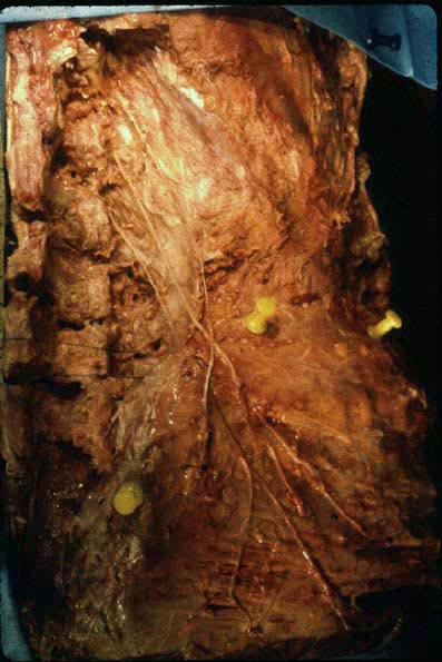
Figure
1 This figure shows the course of the dorsal cutaneous
rami coming from the T11, T12, and L1 roots (superior cluneal nerves). Note
one unusual feature: The ramus coming from the T11 root runs in a medial direction
and crosses over the other two nerves. The pins mark the iliac crest. (Dissection
JY Maigne, with permission from Springer Verlag.10)
The muscle nerves
(both sensory and motor) coming from the thoracolumbar junction also course
caudal. They supply muscles that are about two levels lower down (multifidus),
or which attach to the posterior iliac crest (quadratus lumborum).(2) This innervation
of the more caudal structures by the more cranial level means that a painful
stimulus at the TLJ may give rise to referred pain that is felt very much more
distally, in the lumbar region. This has been confirmed by the injection of
irritant substances into the supraspinous ligament (5) or the facet joints16
at the TLJ, as well as by therapeutic injections at that site, especially when
the needle has penetrated into, or irritated, sensitive tissues.(13) Also, the
pain caused by a crush fracture of T12 or L1 is often felt only in the lower
part of the back.(7)
|
|
The
thoracolumbar junction may give rise to pain |
| |
If pain that is
experimentally produced at the TLJ can be referred to the lower lumbar region,
it is likely that pain from natural causes in that region may equally be referred
distally. There are many potential pain sources at the TLJ: Overall, this level
of the spine is subjected to the same weightbearing stresses as the lumbosacral
junction. In particular, the TLJ is stressed by torquing. While disk herniation
is rare at this level, disk degeneration is frequently seen, and resembles the
lesions encountered at the lumbosacral junction. Vernon-Roberts et al have studied
the T12-L1 disk.(22) They found that concentric tears were more common on the
right than on the left. Radiating tears were seen to be associated with matching
cavitation in the end plate. Rim lesions (anulus tears) were mainly found in
the anterior portion of the anulus, reflecting the stress to which that structure
is exposed. As at the lumbar level, there are synovial folds in the facet joints.(20)
These joints may suffer arthrotic degeneration.(15) The pattern of these joints
may be readily studied in vivo, on CT scans. They typically show a lumbar-type
(sagittal) orientation at T12-L1, and a thoracic (coronal) one at T11-(12).
However, anomalous orientation of the facet joints is frequently seen at T11-12,
with an intermediate pattern (half-way between a lumbar and a thoracic one),
or a asymmetry (articular tropism).(8,15,21) Conventional CT shows that a partially
or completely lumbar pattern limits rotation, and probably transfers the stresses
to T10-11.(8) A slightly asymmetrical orientation of the facet joints may be
a cause of strain, because movement, and especially rotation of the trunk, will
not be smooth.
Fig.
2: Referred low-back pain pattern in the territory of
the dorsal cutaneous branches coming from the thoracolumbar junction (superior
cluneal nerves). The cross marks the "crest point", where the most
medial nerve is in direct contact with the iliac crest at its crossing point.
|
|
The
thoracolumbar junction syndrome |
| |
One of the authors
(RM) has described the clinical features of pain originating at the TLJ.(12,13)
The pain is usually unilateral. It is referred in the posterior dermatomes of
T11, T12, L1 or L2. Thus, the pain is not felt at the site of nociceptive stimulation,
but at a more distal site, at the iliac crest or in the upper part of the buttock.
It may also be referred in the anterior dermatomes, in which case the patient
may complain of pain in the abdominal wall (which may mimic visceral pain),
the groin, or on the outside of the thigh.(14)
Examination
of the painful lumbar region
During the clinical
examination, pain may be aggravated by side-bending in the opposite direction.
This may be due to stretching of the dorsal cutaneous rami by this movement.
Equally, rotation towards the painful side may make the pain worse. Often, the
skin and the subcutaneous tissues will be found to be hypersensitive in the
dermatomes concerned. This hypersensitivity can be demonstrated by a skin-rolling
test. Skin tenderness to pinching and rolling is normally thought to have a
nonorganic basis, if it occurs over a wide and symmetrical area. However, if
the area is localized and confined to one side, pinch-roll tenderness may well
be due to the irritation of a sensory cutaneous nerve.(13,23) The hypersensitivity
observed may be interpreted as an antidromic stimulation of pain nerve fibers
which leads to release of pain-enhancing substances into the skin.(6)
Another typical
clinical feature is the presence, on the painful side, of a tender point on
the posterior iliac crest, at a distance of ca. 7 cm from the midline. This
distance is remarkably constant in different patients. The iliac crest point
is tender, and the sensation elicited by pressure on the point is readily recognized
by the patient as the pain he or she tends to experience. Our anatomical studies
have shown that the iliac crest point is at the site where the most medial of
the superior cluneal nerves crosses over the iliac crest. This nerve comes from
the L1 root in 60% of the cases, and from the L2 root in the remaining 40%.(10)
Clinical examination with fluoroscopic control has shown this iliac crest point
to be consistently at the same site, regardless of the affected level (T11-12,
T12-L1, or L1-L2). This finding may be accounted for by the fact that each facet
joint has a plurisegmental nerve supply,(1) and that pain from one of these
levels may be referred, not only in the corresponding dermatome, but in the
adjacent dermatome(s). In this way, all the superior cluneal nerves would be
sensitized. However, at the point of crossing over the iliac crest, only the
most medial superior cluneal nerve is in direct contact with the bone, whereas
the lateral cluneal nerves are insulated by the subcutaneous fatty tissue. Thus,
tenderness can be more readily elicited by pressure on the medial nerve.
Examination
of the thoracolumbar junction
The TLJ is investigated
with clinical examination, which shows the offending vertebral level; and, if
need be, with an anesthetic block.(12,13) Imaging techniques are of little value,
since there are no specific patterns that could be detected.
For the clinical
examination, the patient is positioned prone, with a pillow under his or her
abdomen; equally, the patient may be placed across the couch. Various maneuvers
are performed to stress the motion segment under examination: extension, by
slowly applied pressure on the spinous processes; torque, by pressure applied
to the sides of the spinous processes; pressure on the facet joints. In a healthy
motion segment, none of these maneuvers should be painful; however, in a segment
that is dysfunctional, from whatever cause, there will be pain. The pain thus
elicited will be on the side of the low back pain.
If the injection
of an anesthetic around the painful facet joint and the corresponding dorsal
ramus relieves the signs and symptoms, this would suggest that the pain is,
indeed, due to a TLJ syndrome. The injection may be made under fluoroscopic
control; or in the clinic or consulting room, using clinical landmarks for guidance.
The technique has been assessed against placebo, in a small series of patients,
and has shown the syndrome to be a genuine entity.(11)
Local injections
and vertebral manipulation constitute the standard treatment of the condition.
|
|
Other
syndromes involving the cutaneous
dorsal rami |
| |
Our understanding
of the conditions involving the dorsal cutaneous rami arising at the thoracolumbar
junction has been further improved by a number of studies.
We performed an
anatomical study, which showed that entrapment may occur at the site where the
most medial cutaneous ramus crosses over the iliac crest, at a distance of ca.
7 cm from the midline.10 The nerve passes through an osseofibrous tunnel, bounded
below by the iliac crest and, above, by the thoracolumbar fascia (which attaches
to the crest). The clinical features observed are similar to those seen in TLJ
syndrome, though, obviously, without any signs of major vertebral dysfunction.
Complete pain relief following the injection of anesthetic into the point on
the iliac crest is diagnostic. Recently, the results of a series of 19 cases
treated with surgical nerve release were published.(9) Also, research done in
Japan has contributed new and interesting data concerning these nerves: The
studies showed that the lower lumbar disks are chiefly innervated by sympathetic
fibers.(17) The nociceptive impulses from these disks are transmitted cranial
via the prevertebral sympathetic trunk; they reach the central nervous system
through anastomoses with the high lumbar roots (L1 and L2). According to the
authors, pain could be referred in the distribution of these roots. This would
account for the way in which sciatic pain may be referred into the groin, and
for the referral in the posterior L1 and L2 dermatomes of some forms of discogenic
pain.18 The authors were able to provide pain relief in patients whose low back
pain was due to one of the two lowest lumbar disks, but referred into the gluteal
region along the cutaneous nerves, using an anesthetic block of the L2 root.
While low back
pain, by definition, is felt in the lumbosacral region, the pain may originate
at the thoracolumbar junction and the sensory nerves arising at that level.
This should be borne in mind in patients whose pain is localized in the thoracolumbar
dermatomes, when pressure on the crest point elicits the pain familiar to the
patient, or if the lumbosacral junction appears not to be responsible for the
patient's problems, or fails to respond to treatment applied at that level.
References
1. Bogduk N. The innervation
of the lumbar spine. Spine 1983;8:286-93
2. Hayashi, N. Tamaki, T. Yamada, H. Experimental study of denervated muscle
atrophy following severance of posterior rami of the lumbar spinal nerves. Spine
1992;17:1361-7.
3. Hirschfeld L. Traité et iconographie du système nerveux et
des organes des sens de l'Homme. 1st ed. Paris: Masson,1866:328-9.
4. Keegan JJ, Garrett FD. The segmental distribution of the cutaneous nerves
in the limbs of man. Anat Rec 1948;102:409-37.
5. Kellgren JH. On the distribution of pain arising from deep structures with
charts of segmental pain areas. Clin Sci Mol Med 1939;4:35-46.
6. Lynn B. Cutaneous hyperalgesia. Br Med Bull 1977;33:103-8.
7. MacNab I. Backache. 1st ed. Baltimore: Williams and Wilkins, 1977:22.
8. Maigne JY, Buy JN, Thomas M, Maigne R. Rotation de la charnière thoraco-lombaire.
Etude tomodensitométrique chez 20 sujets normaux. Annales de Réadaptation
et Médecine Physique 1988;31:239-43.
9. Maigne JY, Doursounian L. Entrapment neuropathy of the medial superior cluneal
nerve. Nineteen cases surgically treated, with a minimum of two years’
follow-up. Spine 1997.
10. Maigne JY, Lazareth JP, Guérin Surville H, Maigne R. The lateral
cutaneous branches of the dorsal rami of the thoracolumbar junction. Surg Radiol
Anat 1989;11:289-93.
11. Maigne R, Le Corre F, Judet H. Premiers résultats d'un traitement
chirurgical de la lombalgie basse rebelle d'origine dorsolombaire. Rev Rhum
Mal Ostéoartic 1979;46:177-83.
12. Maigne R. Low-back pain of thoracolumbar origin. Arch Phys Med Rehabil 1980;61:389-95.
13. Maigne R. Origine dorsolombaire de certaines lombalgies basses : rôle
des articulations interapophysaires et des branches postérieures des
nerfs rachidiens. Rev Rhum Mal Osteoartic 1974;41:781-9.
14. Maigne R. Diagnosis and treatment of pain of vertebral origin. A manual
medicine approach. Baltimore : Williams & Wilkins, 1996:411-7.
15. Malmivaara A, Videman T, Kuosma E, Troup JD. Facet joint orientation, facet
and costovertebral joint osteoarthritis, disc degeneration, vertebral body osteophytosis
and Schmorl’s nodes in the thoracolumbar junctional region of cadaveric
spines. Spine 1987;12:458-63.
16. McCall IW, Park WM, O’Brien JP. Induced pain referral from posterior
lumbar elements in normal subjects. Spine 1979;4:441-6.
17. Nakamura S, Takahashi K, Takahashi Y, Morinaga T, Shimada Y, Moriya H. Origin
of nerves supplying the posterior portion of lumbar intervertebral discs in
rats. Spine 1996;21:917-24.
18. Nakamura S, Takahashi K, Yamagata M, Murakami M,Sugaya H, Sekikawa T, Morinaga
T,Yasuhara K, Moriya H, Takahashi Y. Afferent pathway of low back pain : evaluation
with L2 spinal nerve infiltration. Presented at the annual meeting of the International
Society for the Study of the Lumbar Spine, Helsinski, Finland, 1995.
19. Schwarzer A et al. The relative contribution of the disc and zygapophyseal
joint in chronic low back pain. Spine 1994;19:801-6.
20. Singer KP, Giles LG, Day R. Intra-articular synovial folds of thoracolumbar
junction zygapophyseal joints. The anatomical record 1990;226:147-52.
21. Singer KP, Breidhal PD, Day RE. Variations in zygapophyseal joint orientation
and level of transition at the thoracolumbar junction. Surg Radiol Anat 1988;10:291-5.
22. Vernon-Roberts B, Manthey BA, Fazzalari NL. Three dimensional analysis of
disc pathology. Presented at the annual meeting of the International Society
for the Study of the Lumbar Spine, Helsinski, Finland, 1995.
23. Waddell G, McCulloch JA, Kummel E, Venner RM. Nonorganic physical signs
in low-back pain. Spine 1980;5:117-25.
